Contact : Patrick Aimedieu
The Navier laboratory has an Axiovert Z1 microscope, which includes:
- a part for confocal microscopy. For this application, two laser light sources, with respective wavelengths 488nm and 561nm can be used, coupled with a camera whose acquisition speed is two images per second.
- a part for inverted microscopy coupled to a CCD camera. This microscope also includes a source of white light, transmitted or reflected, and a source of fluorescence in reflection.
This microscope makes it possible to study systems whose characteristic size is of the order of a few microns with very good resolution. It makes it possible, for example, to follow the drying front in an emulsion or to study systems of more or less concentrated particles.
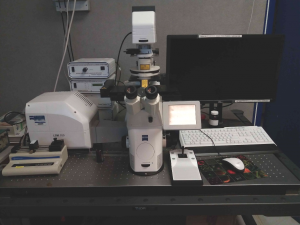
microscope Axiovert Z1
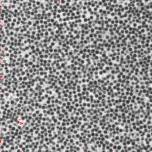
suspension of 2µm silica beads at a volume fraction of 30% and CaCl2 concentration: I = 0.05
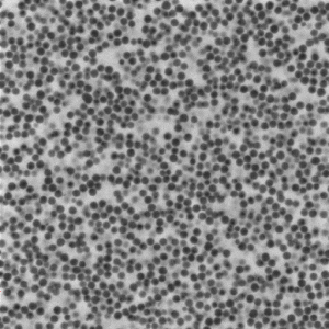
suspension of 2µm silica beads at a volume fraction of 30% and CaCl2 concentration: I = 0.2
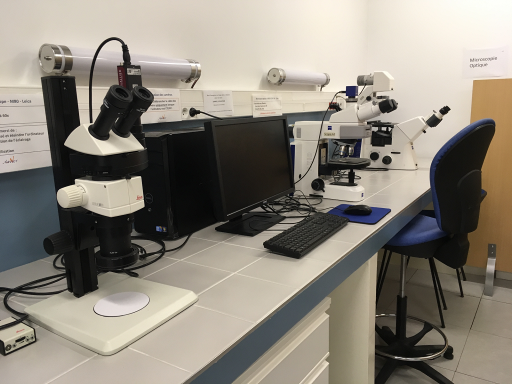
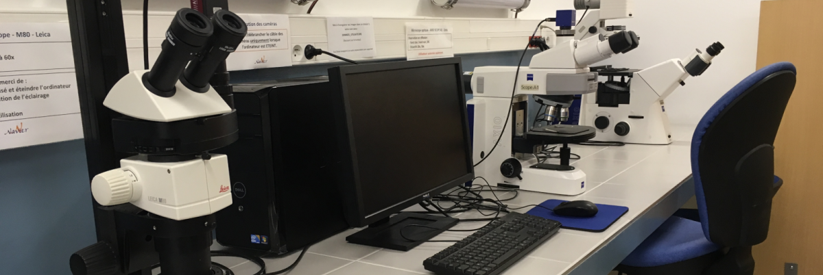
The Navier laboratory also has three other optical microscopes, all equipped with an output to a CCD camera:
- A Leica M80 stereo microscope, with continuous zoom from 7.5x to 60x and LED ring illumination.
- A Zeiss Axio Scope A1 reflected light microscope, allowing observations in bright field, dark field and Differential Interference Contrast (DIC).
- A Zeiss Axio Observer A1 inverted transmitted light microscope, allowing observations in bright field and phase contrast.